Blog
Epithelial Tissue Structure, Types and Function | Free Biology Notes
This article we will discuss about epithelial tissue structure, types of tissue structure and functions of epithelial tissue
- Epithelial tissue forms the lining or covering of internal and external surface of the body
- Epithelium consists of cells arranged in continuous sheets, in either single or multiple layers
Structure of Epithelial Tissue
- The cells in epithelial tissues are very closely packed together and joined
- One surface of epithelial tissue is exposed to either the external environment or the body fluid
- Other surface is attached to tissue by a membrane, which consists of fibres and polysaccharides called basement membrane
- Basement membrane consist of 2 layers
- Basal lamina: Made up of glycoprotein, which is secreted by epithelium cells
- Fibrous lamina: Made up of collagen and reticular fibres are suspended in mucopolysaccharide
- Epithelium has tightly fitted continuous layer of cells. There are specialised junctions present between the cells of the epithelium, that link individual cells called intercellular junction
- Three main types of junctions that are found in epithelial cells
I. Tight junctions
- Plasma membrane of adjacent cells become fused to form tight junction
- Enzymes known as occludin help to fuse the membranes together
- Prevent leakage across tissues
- Found in columnar epithelium
II. Adhering junctions
- The membranes of two adjacent cells aren’t fused together, instead, they adhere to each other in certain places only by actin filaments
- Enzyme known as cadherin helps to keep these filaments together.
- keep the neighbouring tissues well cemented together
III. Gap junctions
- Gap junctions are channels that physically connect adjacent cells
- Protein known as connexins helps to form the pore for a gap junction between the cytoplasm of two adjacent cells
- Facilitate the movement of ions and molecules across the tissue
Classification of epithelial tissue
- On the basis of number of cell layers it has 2 types:- simple epithelium and componund epithelium
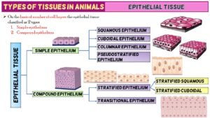
1. Simple Epithelium Tissues
- Composed of single layer of cells
- Functions as lining for body cavities, ducts and tubes
- On the bssis of shape / structural modifications of cells
(a) Simple Squamous epithelial tissue
(b) Simple Cuboidal epithelial tissue
(c) Simple Columnar epithelial tissue
(d) Pseudostratified epithelial tissue
(a) Simple Squamous epithelial tissue
- Consist of a single layer of flattened cells
- Margin of squamous cells are irregular
- Occur as tiles on a floor, also known as pavement epithelium
- Location: Alveoli, Bowman’s capsule, blood vessel, heart, lining of coelom
- Function: Filtration, absorption and secretion
(b) Simple Cuboidal epithelial tissue
- Made up of cube shaped cells.
- It has round nuclei
- Location: Surface of ovary, inner surface of cornea and lens of eye, kidney tubules, salivary and pancreatic ducts
- Functions: secretion and absorption
(c) Simple Columnar epithelial tissue
- Columnar epithelium cells are tall
- They are pillar-like and have elongated nuclei
- Location: Lines stomach, small and large intestine, digestive glands
- Function: Secretion and absorption
(d) Pseudostratified epithelial tissue
- These are simple columnar epithelial cells whose nuclei appear at different heights, giving the misleading impression that the epithelium is stratified when the cells are viewed in cross section
- In this epithelium two types of cells are present
- Long cells
- Extended up to free surface
- Nuclei have oval shape
- Short cells
- Don’t reach the outer surface
- They have rounded nuclei
- Location: Trachea, bronchi, olfactory epithelium, eustachian tube
- Function: Secretion and movement of mucus by ciliary action
2. Compound epithelial tissue
- Composed of multi-layered cells
- Limited role in secretion and absorption
- They cover the dry surface of skin, moist surface of buccal cavity, pharynx, inner lining of ducts of salivary glands & pancreatic ducts
- Provide protection against chemical & mechanical stress
- On the basis of stretching ability it is of 2 types
- Stratified epithelium (Non-stretchable)
- Transitional epithelium (Stretchable)
(a) Stratified epithelium
On the basis of shape of the cells of outermost layer it is of three types
i) Stratified Squamous epithelium
- Innermost layer of cells are cuboidal
- Cells of outermost layer are scale like flat cells
on the basis of presence of keratin protein in the outer most cells this epithelium is of two types
Keratinized stratified squamous epithelium
- Keratin portein is present in scaly cells
- Cells become non nucleated dead cells
- Epidermis of skin, scale, horm, nail, feathers etc.
Non keratinized stratified squamous epithelium
- Keratin protein is absent
- Cells are nucleated and living
- Buccal cavity, inner lining of cheeks, lips, hard plate, pharynx, oesophagus & anal canal
II) STRATIFIED CUBOIDAL EPITHELIUM
- Inner most layer is cuboidal
- Outermost layer of cells are cube like, cells are nucleated & living.
- Secretory duct of sweat glands, mammary glands and sebaceous gland, pancreas, salivary glands
III) STRATIFIED COLUMNAR EPITHELIUM
- Outermost layer is composed of pillar shaped cells, cells are nucleated. On the basis of presence of cilia this epithelium is of 2 types
(1) Ciliated stratified columnar epithelium
Eg. Buccopharyngeal cavity of Frog. Larynx
(2) Non ciliated stratified columnar epithelium. Cilia absent on free end.
Eg. Epiglottis
b) Transitional epithelium
- In a relaxed state it’s appears as stratified cuboidal epithelium but tissue is stretched, cells become flattened, giving the appearance of stratified squamous epithelium
- In this epithelium 4-6 layers of cells are persent
- Inner most layer of cell is composed of cube like cells
- Middle 2-4 layers are composed of pear shaped
- Outermost 1 or 2 layers are of oval shaped cells
- These different shape of cells appears only in resting stage. When this tissue is stretched, all the cells become flattened
- Found in Renal Pelvis, Ureter, Urinary Bladder, Proximal part of male urethra
- Function The transitional epithelium cells stretch readily in order to accommodate fluctuation of volume of the liquid in an organ


 Entertainment1 month ago
Ibomma Bappam: Redefines Telugu Streaming Trend
 Blog1 month ago
[PPT] The living world Class 11 Notes
- Blog1 month ago
[PPT] Human Reproduction Class 12 Notes
- Blog1 month ago
Class 12 Biology Notes Chapter wise PPT
- Blog1 month ago
Iosmirror.cc Apk: Enables Smart Screen Sharing
- Blog1 month ago
PG TRB Botany Study Material PDF Free Download
- Blog1 month ago
Download NEET Biology Study Materials in Tamil
- Blog1 month ago
Class 11 Biology Notes Chapter wise PPT