Blog
Internal Structure of Monocot Leaf Notes | Free Biology Notes
This article we will discuss about Internal Structure of monocot leaf
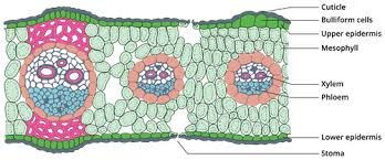
The transverse section of a monocot leaf reveals the following structures
Epidermis
- A monocot leaf is generally isobilateral leaf
- It has upper and lower epidermis
I) Upper epidermis
- Outermost layer present on the upper side of the leaf
- Made up of a single layer of parenchymatous
- Cuticle layer is present on the upper epidermis
- Stomata is present on both upper and lower epidermis
- Some parenchymatous cells on the upper epidermis are large and thin-walled are known as bulliform cells
- Bulliform cells make the leaves curl during water stress and helps to reduce water loss
II) Lower epidermis
- Outermost layer present on the lower side of the leaf
- Single layer, parenchymatous & covered with cuticle
- Number of stomata is equal on both the epidermis
- Stomata is surrounded by dumb bell shaped guard cells
- Function of epidermis: protective layer, exchange of gases and facilitates the transpiration
Mesophyll tissue
- Present between the upper and lower epidermis, there is an entire mass of ground tissue called mesophyll
- Unlike dicot leaf, no differentiation is observed in the mesophyll as palisade and spongy parenchyma
- The cells are made up of parenchyma and are irregularly arranged with intercellular spaces
- These cells contain chloroplasts and can take part in the process of photosynthesis
Vascular bundle
- A large number of vascular bundles are present
- Each vascular bundle consists of xylem and phloem
- Vascular tissues are surrounded by a sheath of cells made up of parenchyma called bundle sheath
- Vascular bundle is conjoint, collateral and closed
- Functions of V.B: conduct water and food materials


 Blog8 months ago
[PPT] Human Reproduction Class 12 Notes
- Blog8 months ago
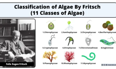
Contribution of Indian Phycologists (4 Famous Algologist)
- Blog8 months ago
PG TRB Botany Study Material PDF Free Download
- Blog8 months ago
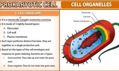
Cell The Unit of Life Complete Notes | Class 11 & NEET Free Notes
 Blog8 months ago
[PPT] The living world Class 11 Notes
- Blog8 months ago
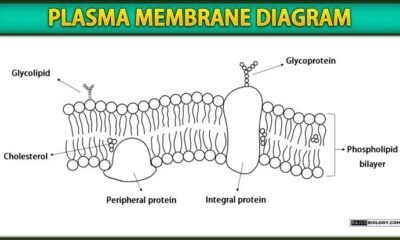
Plasma Membrane Structure and Functions | Free Biology Notes
- Blog8 months ago
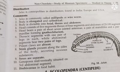
Julus General Characteristics | Free Biology Notes
- Blog8 months ago
Classification of Algae By Fritsch (11 Classes of Algae)