Blog
Cell The Unit of Life Complete Notes | Class 11 & NEET Free Notes
Cell The Unit of Life Class 11 Notes
What is a cell?
- Cell is the building block of all living organisms
Discovery of cells
- In 1665, cell discovered by Robert Hooke
- He actually observed only cell walls
- Hooke coined the term cell
- He wrote his findings in a book Micrographia
- In 1672, Anton Von Leuwenhoek first saw and described live cell
- Robert Brown later discovered the nucleus
Cell theory
- In 1839, Schleiden and Schwann formulated the cell theory
- All living organisms are made up of cells
- In 1855, Cell theory was further modified by Virchow
- All cells arise from pre existing cells by cell division (Omnis cellula e cellula)
Size of Cells
- Smallest Cell: Mycoplasma 0.3 μm
- Largest Cell: Egg of Ostrich 15 cm
- Longest Cell: Nerve cells 100 cm
- Human red blood cell: 7μm
- Bacterial Cell: 3 to 5 μm
Shape of Cells
- Shape of cell may very with the function they perform
- They may disc-like, Polygonal, Columnar, Cuboids and thread like
Types of cells
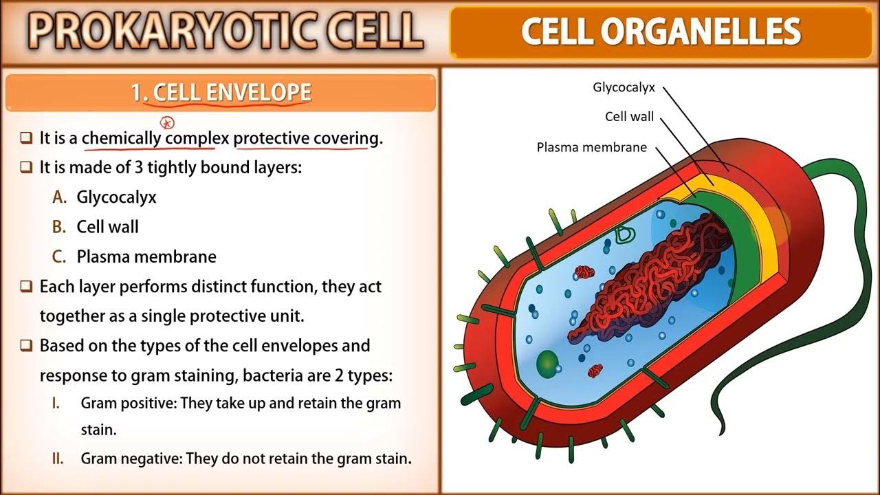
I) Prokaryotic cell
- Cells without nucleus
- E.g. Bacteria
II) Eukaryotic cell
- Cells with nucleus
- E.g. Plant & Animal cells
I) Prokaryotic cell
- Prokaryotic cells are single celled microorganisms
- Prokaryotes include all bacteria
- All the reactions occur within the cytoplasm
- They can be free living or can be found within the gut of other organisms
Characteristics of prokaryotic cell
- Do not have a true nucleus
- Membrane bound organelles absent
- Mitochondria, Golgi bodies, chloroplast, and lysosomes are absent
- Plasma membrane acts as the mitochondrial membrane carrying respiratory enzymes
- Cell wall is peptidoglycan
- Some may have flagella for movement
- Smaller & less complex than eukaryotes
Prokaryotic cell structure
- Capsule: Outer protective covering, Helps in moisture retention and protection
- Cell Wall: Outer layer of the cell. Its gives shape
- Cell Membrane: This layer surrounds the cytoplasm. regulates the entry and exit of substances in the cells
- Pili: Hair-like outgrowths that attach to the surface of other bacterial cells
- Flagella: Long rope like structures, help in the locomotion
- Cytoplasm: Cytoplasm is mainly composed of enzymes, salts, cell organelles and is a gel-like component
- Ribosomes: Involved in protein synthesis
- Plasmids: Plasmids are non-chromosomal DNA structures
- Nucleoid Region: It is the region in the cytoplasm where the genetic material is present
II) Eukaryotic cell
- Eukaryotes include all the protists, plants, animals and fungi
- Eukaryotic cells have a true nucleus
- They have membrane-bound organelles
- All eukaryotic cells are not identical
- Typical cell consists of
- Cell Wall
- Plasma Membrane
- Cytoplasm
- Nucleus
1. Cell wall
- Discovered by Robert Hooke in 1665
- Cell wall non-living rigid structure
- Found in plants, fungi, prokaryotes and protists
- Composition of cell wall varies in different groups
Composition of cell wall
Prokaryotes Cell Wall
- Cell wall is made up of peptidoglycon
- N-Acetyl Muramic acid and N-Acetyl Glucosamine
Fungi Cell Wall
- Cell wall is made up of chitin
- N-Acetyl Glucosamine
Plant Cell Wall
- Cellulose – D glucose units
- Hemicellulose – Arabinose, mannose, xylose & galactose
- Pectin – Galactose, galacturonic acid and arabinose
- H2O, Lipids and proteins
- Extra substance: Cutin, Subarin & Lignin
Structure of cell wall
- Cell wall is made up of 4 layers
- Middle lamella, primary, secondary and tertiary wall
I) Middle Lamella
- Cementing layer between the cells
- It is made up of Ca & Mg pectates
II) Primary Cell Wall
- Found in growing cell
- It has high hemicelluloses & less cellulose content
III) Secondary Cell Wall
- Found in Mature cell
- It has high cellulose & less hemicelluloses content
IV) Tertiary Cell Wall
- Laid down on secondary wall
- Found in tracheids of gymnosperms
- Cell wall is not uniform in thickness throughout
- Certain places cell wall are not laid down, Such places are called pits
Pits are of two types
I) Simple Pit
- Pit chamber is uniform in diameter
- Found in angiosperms
II) Bordered pit
- Pit chamber is flask shaped
- Found in gymnosperms
- Number of plasmodesmata or cytoplasmic strands are present in pit
Functions of cell wall
- Maintains shape of the cells
- Protects cell from mechanical injury
- Allows materials to pass in and out of the cell
- Prevents undue expansion of cell when water enters by osmosis
Plasma membrane
- Plasma membrane is thin, elastic semi permeable living membrane that serves as boundary for the cytoplasm
- Plasma membrane was coined by nageli in 1855
- Cell membrane or plasmalemma
- Made up of protein and phospholipids
- Selectively permeable in nature
- Dynamic membrane
Structure of plasma membrane
- Three important models explaining the structure of plasma membrane are
- Sandwiched model
- Unit membrane model
- Fluid mosaic model
1) Sandwiched model
- In 1935, Proposed by Davson & Danielli
- Oldest model on the structure of plasma membrane
- Plasma membrane is made up of three layers
- Outer protein layer, Middle lipid layer & inner protein layer
- Proteins are alpha globular
- Lipids are amphipathic
- Lipids arranged in bilayer in such a manner that tails face each others
- Van der vaal force helping to 2 layers stay together
- Proteins on either side of phospholipids bilayers
2) Unit membrane model
- In 1959, Proposed by Robertson
- Plasma membrane is made up of three layers
- Outer protein layer, Middle lipid layer & inner protein layer
- Proteins are beta fibers
- Thickness of plasma membrane is 7.5nm (75Å)
- Lipids are amphipathic
- Lipids arranged in bilayer in such a manner that tails face each others
- Van der vaal force helping to 2 layers stay together
- Proteins on either side of phospholipids bilayers
3) Fluid mosaic model
- In 1972, Proposed by Singer & Nicolson
- Made up of protein and phospholipids
- Proteins are alpha globular & two types
I) Extrinsic Proteins
- Arranged on surface of lipid heads
- Loosely attached
II) Intrinsic Proteins
- Deeply embedded
- Tightly bound to lipids
- Phospholipids have two types of movements
I) Transition
- Movement of phospholipids molecules in the same layer
II) Flip Flop
- Movement of phospholipids molecules between two layers
- Protein iceberg in a sea of phospholipids
Function of Plasma Membrane
- Mechanical support: It gives shape to the cell and protects all cell contents
- Exchange of materials: Regulate the exchange of materials. It allows need materials to enter the cell. It Send out unwanted materials from the cell
- Endocytosis: Engulfing of food or foreign particles through the plasma membrane. Endocytosis differentiated into two types
I) Phagocytosis
- Engulfing of solid particles through the plasma membrane
- g. Amoeba & WBC
II) Pinocytosis
- Engulfing of fluid particles through the plasma membrane
- g. Epithelial cells of intestine
Exocytosis: Process of exudating secretary products from the cells to outside of the cytoplasm E.g. Pancreatic cells
Transport through Plasma Membrane
I) Passive Transport
- No need ATP
- Movement of substance according to concentration gradient
- Higher concentration to lower concentration
Osmosis: Movement of solvent through selectively permeable membrane
Diffusion: Movement of molecules through selectively permeable membrane
Facilitated diffusion: Movement of molecules through transport protein in plasma membrane
Ii) Active Transport
- Need ATP
- Movement of substance against to concentration gradient
- Lower concentration to higher concentration
- Na+/K+ pump
- Ca pump
Specialization of Plasma Membrane
- Plasma membrane shows some specialized structures
- To perform some additional functions plasma membrane shows some changes
Free surface modification
Microvilli
- Minute finger like projections arising from surface of certain cells
- Single cell contains more than 3000 microvilli
- Found in epithelial cells of intestine, kidney tubules, gall bladder and hepatic cells
- Function: increase the surface area for absorption
Junctional complex
Inter digitations
- Plasma membrane of adjacent cells project into cytoplasm as finger-like projections
- Found in lymph nodes and lymphoid tissues
- Function: increase surface area for exchange of substance between the cells
Tight junction
- Plasma membrane of adjacent cells fuses by extrinsic proteins
- Found in brain cells, gall bladder and intestinal cells
- Function: adhesion
Gap junction
- Gap junction is channel through two cell membranes across the intercellular space between two adjacent cells
- Found in cardiac muscles
- Function: conduct electrical signals & passage of ions, sugar, vitamin and metabolites
Desmosomes
- Desmosomes are thickened areas of plasma membrane of two adjacent cells
- Found in cardiac muscle and skin cells
- Function: help to glue the cells together
Plasmodesmata
- Cytoplasm of adjacent cell connected with cytoplasmic strands
- Found in plant cells
- Function: exchange of materials between two cells
Cytoplasm
- Present between plasma membrane and nuclear membrane
- Cytoplasm is jelly like substance
- Cytoplasm also known as cytosol or cytoplasmic matrix
- Autonomic movement of cytoplasm in a cell is called cytoplasmic streaming or cyclosis
- Cytoplasmic structures comprises 3 groups
- Cytoplasmic Matrix
- Cytoplasmic Organelles
- Cytoplasmic inclusions
- Cytoplasmic matrix exists in two states
Gel or plasmagel
- Thick semi solid colloidal solution
Sol or plasmasol
- Liquid colloidal solution
- Cyclosis only occur in sol
Functions of Cytoplasm
- Site for cell organelles and cell inclusions
- Site for metabolic reactions
- Pseudopodia formation
Cytoplasmic Organelles
Endoplasmic Reticulum
- In 1897, observed by garnier and named it as ergastoplasm
- In 1945, first described by porter, claude and fullman
- In 1953, term given by porter
- Present in all eukaryotes
Components of Endoplasmic Reticulum
I) Cisternae
- Formed by projection of nuclear membrane
- They bear ribosomes on the surface
- Contain ribophorin that bind the ribosomes
II) Vesicles
- They are oval or rounded, vacuole like element
- Many vesicles are free in the cytoplasm
III) Tubules
- They are tube like extensions
- Tubules are irregular and branched
- Free of ribosomes
Types of Endoplasmic Reticulum
I) Rough Endoplasmic Reticulum
- Ribosomes binds with help of ribophorin protein
- Mainly composed of cisternae
- Abundantly occurs in cell which are engaged in protein synthesis and secretion
- Mass of RER in cyton of nueron is called missals granules
- G. Liver, pancreas and goblet cells
II) Smooth Endoplasmic Reticulum
- Ribosomes and ribophorins are absent
- Mainly composed of tubules
- Occurs especially in those cells which are almost inactive in protein synthesis
- It is well developed in cells that synthesis lipids
- Mass of SER in retinal cell is called myeloid bodies
Functions of Endoplasmic Reticulum
- It gives mechanical support so also called cytoskeleton of cell
- Intracellular exchange: transport of materials in cytoplasm from one place to another through the ER
- Provides site for protein synthesis, because rough ER has ribosome’s
- Lipids synthesized by smooth ER
- Detoxification of drugs
Golgi Complex
- Observed by George
- Morphological structure given by Camillo Golgi
- Ultra structure given by Dalton
- Golgi complex also known as Dalton complex, lipochondria, dictyosome and golgisome
- Golgi bodies located in the cytoplasm
- Animal cells contain one Golgi complex but plants contain more than 100
- Golgi body size is bigger in secretary cells
- Clear cytoplasm area around Golgi body is called zone of exclusion
Structure of Golgi complex
- Golgi complex is made up of three parts
I) cisternae
- These are slightly curved & sacs like structure
- Many cristenae are arranged parallel to each other
- Convex surface towards nucleus is called cis face or forming face
- Concave surface towards cell membrane is called trans face or maturing face
- Cis and trans faces are inter connected
II) Vacuole
- Spherical and bigger in size
- Found in closer to concave surface
III) Vesicles
- They are spherical sac like structure
- Found in closer to convex surface
Function of Golgi complex
Secretion
- Major function of Golgi body is secretion
- Secretion involve three steps
- Golgi body receives the materials from ER
- These materials are chemically modified
- After modifications materials are packed in vesicles. These vesicles are pinched off from trans face and discharged outside the cell
Middle man of cell
All macromolecules which are to be sent out side cell, move through the Golgi body So Golgi body termed as director of macromolecular traffic in cell
Formation of lysosome
- Golgi body involved in formation of primary lysosomes
- ER buds off small vesicles containing hydrolases
- This vesicle transfer to Golgi complex
- Cisternae of Golgi complex intern bud off small vesicles called primary lysosomes
Lysosome
- Discovered by christian de duve in 1955
- In 1956, novikoff coined the term lysosome
- Found in all eukaryotic cells
- In animals, lysosomes are abundant in leucocytes, macrophages and similar cells with phagocytic activity
- Lysosome are small, spherical and single membrane bounded and vesicular organelles of the cytoplasm
- They contain more than 50 enzymes, important enzymes are lipases, proteases and carbohydrases
- Lysosomes are highly polymorphic cell organelles
Types of Lysosomes
I) Primary lysosomes
- These are store acid hydrolases in inactive form
- These are new newly formed lysosome
II) Secondary lysosomes
- Formed by fusion of primary lysosomes and pinosomes or phagosomes
- It’s have an active acid hydrolases
III) Tertiary lysosomes
- Lysosomes containing undigested material
- These are eliminated by exocytosis
IV) Auto lysosomes
- Lysosomes containing cell organells to be digested are autophagosomes
- These are called suicidal bags
Functions of Lysosome
Interacellular digestion
Heterophage: This is digestion of foreign materials received in cell by phagocytosis or pinocytosis
Autophagy: Digestion of old or dead cell organelles. Autophage also takes place during starvation of cell
Cellular Digestion or Autolysis
- Sometimes all Lysosomes of a cell burst to dissolve the cell completely
- Old cells are removed by autolysis
- Unwanted organs are destroyed by autolysis
GERL System
- It is also known as endomembrane system
- It is a group of organelles in eukaryotic cells that works together to modify, package and transport lipids and proteins
- GERL system consist for golgi apparatus, endoplasmic reticulum and lysosome
Function of GERL System
- Endocytosis of outer substances
- Exocytosis of cellular substances
Ribosomes
- Observed by Claude in 1941 & named as microsomes
- Palade coined the term ribosome
- Ribosomes found in all cells
- Ribosomes are site of protein synthesis in a cell hence called protein factory
- They are located in cytoplasm or attached to ER
- They also present inside some cell organelles
Struture of Ribosomes
- Ribosomes are spherical bodies
- Ribosomes consists of two sub units
- They are large sub units and small sub units
- Sub units occur separately in the cytoplasm
- They join together to form ribosomes only at the time of protein synthesis
- During protein synthesis many ribosomes line up and join mrna chain. Such a string called polyribosome
- Small sub unit hold mrna
- Large sub units have three slots
- A site: trna first binds to mrna
- P site: amino acid added to polypeptide chain
- E site: exit site
Types of Ribosomes
- I) 70s ribosomes
- Found in prokaryotic cells
- Smaller in size and it has sedimentation coefficient is 70s
- 70s ribosomes is made up of two sub units
- Large 50S and small 30S
II) 80s ribosomes
- Found in eukaryotic cells
- Larger in size and it has sedimentation coefficient is 80s
- 80s ribosomes is made up of two sub units
- Large 60S and small 40S
Function of Ribosomes
- Protein synthesis
- Newly formed polypeptide is protected from degradation by cytoplasmic enzymes
Mitochondria
- Discovered by kolliker
- Mitochondria term given by benda
- Power house of cells or ATP mill in cell
- Number of mitochondria
- Depends upon the type and physiological activity of cell
- One per cell in chlorella and chlamydomonas
- 50000 per cell in amoeba
- 5 lacks per cell in flight muscle of insects
- Size of mitochondria
- Smallest mitochondria is seen in yeast (1 μm)
- Largest mitochondria is seen in oocytes of amphibian (20-40μm)
Structure of Mitochondria
- Mitochondria is covered by double membrane
- Two membranes are separated by a space called perimitochondrial space
- The central space filled with mitochondrial matrix
- Inner membrane shows a number of infoldings are known as cristae
- In metabolically active mitochrondria number of cristae is higher
- The inner surface is covered by inner membrane subunits, also called F1 subunits or mitochondrial particles or oxysomes
- Oxysome consists of 3 parts – head, stalk and base
- These particles contain ATP synthetase, which are involved in ATP synthesis
- Matrix in mitochondria
- Pale yellow color
- Contain enzymes for krebs cycle
- Contain divalent ions like mg 2+, mn 2+, fe 2+
- DNA and ribosomes
Function of Mitochondria
- Mitochondria are site of aerobic reparation and ATP production
Plastids
- Plastids are pigment containing cell organelle found in photosynthetic cells
- It is double membrane organelle
- Discovered by haeckel (1865)
- Plastids classified into two groups
- Leucoplast
- Chromoplast
Leucoplast
- Leucoplasts are colorless plastids
- Oval, spherical rod like or filamentous
- Leucoplasts act as storage organelles
- Don’t have pigments and granum
- Found in seeds, tubers and rhizomes
- Classified into three types on the basis of the material stored
- Amyloplast: Stores starch
- Aleuroplast: Store protein
- Elaioplast: Stores droplets of oil or fats
Chromoplast
- Chromoplasts are colored plastids
- It is responsible for colors in flowers, ripening fruits, autumn leaves and some root like carrot
- Chromoplast again classified into following groups
- Chloroplast
- They contain chlorophyll pigment
- Found in green parts of plants
- Phaeoplast
- They contain Fucoxanthin pigment
- Found in Brown algae and diatoms
- Rhodoplasts
- They contain phycoerythnin pigment
- Found in Red algae
Chloroplast
- Discovered by anton von leuwenhoek
- Chloroplast acts as factory for the synthesis of sugars in autotrophic eukaryotes
- No of chloroplast varies
- Chlamydomonas & ulothrix contain 1 per cell
- Higher plants contain 20-40 per cell
- Chara contain 500 per cell
- Size & shape of chloroplast varies
- Chlamydomonas have cup shaped
- Spirogyra have spiral or ribbon shaped
- Ulothrix have girdle shaped
- Zygnema have star shaped
Structure of Chloroplast
- Chloroplast is covered with a double membrane
- Two membranes are separated by a space known as periplastidial space
- Inner content of chloroplast is matrix
- In stoma of flatted sacs are present called thylakoids
- Thylakoids arranged in stacks like piles of coins called grana
- Each chloroplast contains about 20-100 granum
- Each granum is composed of 10-100 thylakoids
- Membrane of thylakoid contain number of minute particles called quantosomes
- Quantasome contains of about 250-400 molecules of various pigments
- Like Chlorophylls, Carotenoids, Xanthophylls etc
- Stoma of chloroplast contains enzymes for the synthesis of carbohydrate and protein
- Stoma contain small double stranded circular DNA molecules
Functions of Chloroplast
- Photosynthesis: Trap light energy of sun and transform it into chemical energy
- Starch Storage: Temporarily store starch during day time
- Protein synthesis: They have own DNA and ribosomes and they synthesis certain amount of proteins
- Oxygen supply: Utilize carbon dioxide for photosynthesis and release oxygen
Cilia and flagella
Cilia
- Cilia are small hair like structure
- Around 5-10 μm in length
- Only found in eukaryotic cells
- Occur throughout the cell
- Involved in processes like locomotion and feeding
- Types: kinocilia & steriocilia
Flagella
- Flagella are long whip like structure
- Around 150 μm in length
- Found in both cells
- Occur at one end of a cell
- Involved in locomotion
- Types: whiplash & tinsel
Structure of cilia & flagella
- Made up of plasma membrane and axoneme
- In axoneme have 9 peripheral doublet microtubules and 2 central singlet microtubules
- This arrangement is called 9 + 2
- The central tubules are connected by bridges and enclosed by central sheath
- Central tubules also connected to peripheral doublets by radial spoke
- Peripheral doublets interconnected by linkers
Centrosomes
- Centrosome term given by theodar boveri
- Centrosome containing two centrioles
- Centrioles are cylindrical and made up of micro tubular protein
- Centrioles occur in nearly all animal cells and motile plant cells
- These are also called diplosomes
- Its look like cart wheel
Structure of Centriole
- Centriole contain 9 peripheral microtubule
- Microtubule are absent in the centre
- The arrangement is therefore called 9+0
- Each microtubule in a triplet. These triplets are linked with the help of A-C linker
- Central part of centriole is proteinaceous and called hub
- Hub connected with peripheral triplets by radial spokes
- Centrioles are self duplicating units
Functions of Centriole
- In animal cells centrioles play important role in cell division by arranging spindle fibers
- Centrioles form the basal body of cilia or flagella
Vacuoles
- Discovered by antonie van leeuwenhoek
- Vacuole is single membrane bound space found in cytoplasm
- Vacuole is not an air filled cavity, rather it is filled with sap
- Sap contain water, excretory product and other materials
- In plant cells the vacuoles can occupy up to 90 per cent of the volume of cell
- In animal cells small sized vacuoles may be found, numbering few to many
- Vacuole may retain ions and other materials against concentration gradient
Types of vacuoles
Sap vacuoles
- Bubble like sac
- Contain cell sap
- Contain water soluble vacuoles pigments like anthocyanin and anthoxanthine
- Present in plant cell
Contractile vacuoles
- Found in freshwater protozoan
- Helps in osmoregulation and excretion
Food vacuole
- Occur in protozoan, protists and lower animals
- Contain digestive enzymes
Air vacuole
- Found in aquatic plants
- Store gases, regulate buoyancy of cell, provide mechanical support etc
Functions of vacuole
- Removing unwanted structural debris
- Isolating materials that might be harmful
- Containing waste products
- Exporting unwanted substances from the cell
Microbodies
- Discovered by rhodin in 1954
- Found in all eukaryotic cells
- Microbodies are minute, simple, roughly spherical sacs bounded by a single unit membrane
- Microbodies bud off from the rough endoplasmic reticulum
- Microbodies are of two main types: peroxisomes and glyoxysomes
Types of Microbodies
Peroxisomes
- Found in both plant and animal cells
- Peroxisomes contain catalae enzymes
- Its help in peroxide metabolism
- Catalase degrade the H2O2 into water and oxygen
Glyoxysomes
- Glyoxysomes occurs only in plants
- Especially in fatty seeds
- Help in glyoxylate acid cycle
- This cycle convert fats into carbohydrates
Cytoskeleton
- Cytoskeleton is network of filamentous proteins in cytoplasm
- Cytoskeleton is made up of microtubules and microfilaments
Microtubules
- Microtubules are hollow rod like structures
- Made up of tubule protein
- Microtubules are thickest cytoskeleton component
- Maintain cell shape & help cell division
Microfilaments
- Microfilaments are solid rod like structures
- Made up of 2 twisted strands of the actin protein
- Microfilaments are thinnest cytoskeleton component
- Microfilaments serve a number of functions: support, movement of villi, movement of plasma membrane and formation of spindle
Cell inclusions
- All non living things in cytoplasm are known as cell inclusions
- They are formed during metabolism of the cell
- Three types of cell inclusions
- Reserve food substance
- Secretary substance
- Excretory substance
Types of cell inclusions
Reserve food: They occur in the form of starch, glycogen, fat droplets and aleurone grains
Secretory products: Many substances secreted by special glands and organs
Excretory products: Several chemical substances which are of no use to produce during metabolic reactions. These waste products are called excretory products
Nucleus
- Discovered by robert brown in 1831
- Study of nucleus is known as karyology
- Nucleus is the control centre of the cell for cell metabolism and reproduction
- Nucleus present in all eukaryotic cells
- Number of nuclei: usually one but may be more than 1 as in few protists
- Position of nuclei: centric in animal cells, ecentric in plant cells and basal in columnar cells
Structure of nucleus
- Ultra structure of the nucleus consists of following components
- Nuclear membrane
- Nucleoplasm
- Nucleolus
- Chromatin
I) Nuclear membrane
- Nuclear contents are set apart from the cytoplasm by a double membrane called the nuclear envelope
- Outer membrane is connected with endoplasmic reticulum and its outer surface may contain ribosomes
- Inner membrane is smooth and its devoid of ribosomes
- Nuclear membrane contain many pores called nuclear pores
- Nuclear pore surrounded by circular structure known as annulus
- Nuclear pore + annulus = nuclear pore complex
- Exchange of materials occurs through the nuclear pores
Nuclear envelope functions
- Maintains the shape of the nucleus
- Regulates the flow of materials into and out of the nucleus
II) Nucleoplasm
- Inner to the nuclear membrane, the nucleus filled with semi solid substance is called nucleoplasm
- Also known as karyolymph or matrix or nuclear sap
- Nucleoplasm contains nucleotides, enzymes and metal ions
Nucleoplasm functions
- It is the seat of synthesis of DNA and rnas
- It provides turgidity to nucleus
III) Nucleolus
- Discovered by fontana in 1781
- Term was coined by bowman in 1840
- The nucleolus is large, spherical and dense body seen inside the nucleus
- Nucleolus is composed of rrna and phosphoproteis
Functions of nucleolus
- Centre for the formation of mrna
- Stores mrna
IV) Chromatin
- Nucleoplasm contain many tread like, elongated and coiled structure known as chromatin fibers
- During cell division, the chromatin fibers become thick strand like structures known as chromosomes
- Chromosomes contain DNA and nucleoproteins
Chromatin has two types
Euchromatin
- Active part of chromosomes
- Contain functional unit is called gene
Heterochromatin
- Inactive part of chromosomes
- Contain small amount of DNA and large amount of RNA
Functions of Nucleous
- Control all the activities of the cells
- It play significant role in heredity
- Nucleus contain genetic code for protein synthesis
- Synthesis of ribosomal RNA takes place in nucleus
Chromosomes
- Discovered by Holfmeister in 1848
- Term was coined by Waldeyer in 1888
- Chromosome is highly condensed form of the chromatin
- No of chromosomes varies from species to species
- Lower number of chromosomes found in Ascaris sp. (2)
- Maximum number of chromosomes found in radiolarian (1700)
Structure of chromosome
Pellicle
- It is the outer envelope
- It is very thin
Matrix
- Ground substance of chromosome which contains the chromonemata
- Matrix is enclosed in a sheath which is known as pellicle
- Matrix and pellicle are non-genetic materials
Chromatid
- Two longitudinal subunits of chromosome is called chromatid
- Both chromatids joined together by common centromere
- Chromosomes have two chromatis in prophase and metaphase
- Chromosomes have single chromatid in anaphase and telophase
- Each chromatid is consist of DNA associated with histone proteins
Centromere
- Sister chromatids are joined by the centromere
- The position of centromere differs in chromosomes
- The centromere is called primary constriction
- Centromere divides chromosome into two parts, shorter arm is known as ‘p’ arm and the longer arm is known as ‘q’ arm
- Centromere contains a disc-shaped kinetochore
- Kinetochore is actual site of attachment of spindles to chromosomes during cell division
Secondary constriction
- Constricted region other than that of centromere is called secondary constriction
- Satellite: chromosome segment separated from the main body of chromosome by one secondary constriction
- Chromosome with secondary constriction is referred to as satellite chromosome or sat-chromosome
Telomere
- Terminal region of a chromosome on either side
- Each chromosome has two telomeres
- Telomeres are polar, which prevents the fusion of chromosomal segments
Chromonema
- Thread like coiled structures
- Embedded in the matrix
- Chromonemata formed of a double helix of DNA
Classification of chromosomes
Basis of location of centromere
Telocentric
- Centromere present on the terminal position
- No ‘p’ arm (short arm) present
- Telomeric chromosomes are not found in humans
Acrocentric
- Centromere occupying a sub-terminal position
- One arm is very long and the other is very short
- Chromosomes 13 – 15, 21, 22, Y
Submetacentric
- Centromere slightly away from the mid-point
- Two arms are unequal
- Chromosomes 2, 4 – 12, 17, 18, X
Metacentric
- Centromere lies in the middle of chromosome
- Two arms are almost equal
- Found in chromosomes 1, 3, 16, 19, 20
Function of Chromosomes
- Genetic code storage: Chromosome contains the genetic material that is required by the organism to develop and grow. Genes are those sections of the DNA which code for specific proteins required by the cell for its proper functioning
- Sex determination: Humans have 23 pairs of chromosomes out of which one pair is the sex chromosome. Females have two X chromosomes and males have one X and one Y chromosome. Sex of the child is determined by the chromosome passed down by the male
- Formation of proteins and storage: Chromosomes direct the sequences of proteins formed in our body and also maintain the order of DNA.
Download PDF Study Material


 Entertainment1 month ago
Ibomma Bappam: Redefines Telugu Streaming Trend
 Blog1 month ago
[PPT] The living world Class 11 Notes
- Blog1 month ago
[PPT] Human Reproduction Class 12 Notes
- Blog1 month ago
Class 12 Biology Notes Chapter wise PPT
- Blog1 month ago
Iosmirror.cc Apk: Enables Smart Screen Sharing
- Blog1 month ago
PG TRB Botany Study Material PDF Free Download
- Blog1 month ago
Download NEET Biology Study Materials in Tamil
- Blog1 month ago
Class 11 Biology Notes Chapter wise PPT