Table of Contents
Connective tissue: Definition, Structure, Types and Function
In this article we will discuss about the connective tissue: – Definition, Characteristics, Structure, Types and Function of connective tissue
Definition of Connective tissue
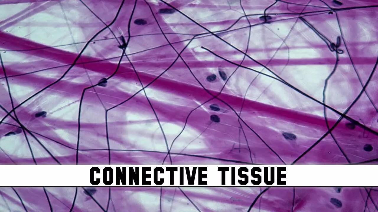
Connective tissue may be defined as an animal tissue that supports, connects, and surrounds organs and other body parts. It is the most abundant and widely distributed tissue in the body.
Characteristics of connective tissue
- It’s composed of the matrix and fibers
- Connective tissues bind one tissue to another
- It’s not present on free surfaces or body cavities
- They provide support to cartilages and bones
- They connect various body organs
Structure of connective tissue
- It’s is composed of two components: –
- Different types of cells
- Matrix
- Variations in the composition and amount of the components are responsible for the typing of connective tissue
i) Cells of connective tissue
- The cells of connective tissue are living
- They are responsible for secreting large amounts of intercellular ground substance
Fibroblast cells
- Largest cells and maximum in number
- Cytoplasm branched so they appear irregular in shape
- They are chief matrix producing cell
- Function: produce fibres and secrete matrix
Macrophages
- Modified monocytes cells
- They are phagocytic in nature and also called scavenger cells
- Function: destroy bacteria, virus and dead or damaged cells of connective tissue
Mast cells
- Mast cells are oval cells with secretory granules in the cytoplasm
- These cells have s shaped nucleus
- Function: – storing histamine (vasodilator), serotonin (vasoconstrictor), heparin (anticoagulant) and secrete matrix
Adipose cells
- Oval shaped cells which store fat
- Function: – store fat
Lymphocytes
- Amoeboidal in shape with a large nucleus
- Function: produce, transport & secretes antibodies
Plasma cells
- Amoeboidal in shape with rounded nucleus
- Function: produce, secrete & transport antibodies
ii) Matrix of connective tissue
- It fills the space between the cells
- It has 2 major components: –
- Ground substance
- Fibres
i) Ground substance
- It’s composed of mucopolysaccharide and water
- Transparent material with the properties of a viscous
- Extracellular material which serves as a diffusion medium in the spaces around the cells and fibers
- It plays major role in determining the physical nature of connective tissue
ii) Fibers
- Fibers are elongated proteins secreted by fibroblast cells
- Fibers fond in matrix between the connective tissue cells
- Function is strengthened and support the connective tissues
- There are 3 different types of fibres
Collagen fibers (white fibres)
- They are white in colour composed of collagen protein
- Very strong and resist tension, allow flexibility
- Found in bone and cartilage
Elastic fibres (yellow fibres)
- They are yellow in colour composed of elastin protein
- Possess elasticity, ability to return to its original shape after being stretched
- Found in skin and wall of blood vessels
Reticular fibres
- They are composed of reticulin protein
- Highly branched fibres which always form dense network
- Found in lymphoid organs like spleen or lymph nodes
Classification of connective tissue
- The primary elements of connective tissue include a ground substance, fibers and cells.
- Based on structural characteristics or major component of cell, connective tissues classified into three main groups
- Loose connective tissue
- Dense connective tissue
- Specialized connective tissue
i) Loose connective tissue
- Distributed throughout the body as a binding and packing material
- Cells and fibres are loosely arranged in a matrix
- It’s contains more ground substance and fewer cells and protein fibers
- There are three types of loose connective tissue:
- Areolar connective tissue
- Adipose connective tissue
- Reticular connective tissue
a) Areolar tissue
- Widely distributed connective tissues
- Consists of cells, fibers and ground substances
- The most abundant cells are fibroblasts and predominant fibers in the matrix are collagen fibers
- It serves as a support framework for epithelium
- Located under the skin, covering of the blood vessels, esophagus and other organs
b) Adipose tissue
- They are fat-filled tissues that have adipocytes
- They store energy, conserve body heat, fills space in the body parts and guards many organs and shape up the body
- Located under the skin, kidneys, eyes and heart
c) Reticular connective tissue
- It’s containing network of reticular fibers, on which fibroblast and leukocytes are suspended
- It has very little ground substance
- Mainly present in hematopoietic system, like spleen, lymph nodes, bonemarrow.
- They form stroma of organs, filters and removes worn-out blood cells in spleen and microbes in lymph nodes
ii) Dense connective tissue
- It’s consists of densely packed fibers with relatively little space between the fibers
- It has proportionately high protein fiber than ground substance
- Abundance of collagen fibers also known as collagenous connective tissue
- Further divided into three categories.
- Dense regular connective tissue
- Dense irregular connective tissue
- Elastic connective tissue.
a. Dense regular connective tissue
Show regular pattern of fibres
Lots of collagen fibres that are parallel to one another and are packed tightly
Examples:- Tendons: connect muscle to bone and Ligaments: connect bone to bone
b. Dense irregular connective tissue
- Show irregular pattern of fibres
- They are pretty strong and provides protection to organs from injury
- Located in the dermis of the skin, capsules around spleen and liver, the fibrous sheath around bones, and other organs.
c. Elastic connective tissue
- It’s composed primarily of elastic fibers and yellowish in color
- They can be stretched to one and a half times their original lengths and will snap back to their original size
- Found in the walls of large arteries, in the vocal cords, and in the trachea and bronchial tubes of the lungs.
iii) Special connective tissue
- Made up of different tissues with specialized cells and unique ground substances
- Some of these tissues are solid and strong, while others are fluid and flexible
- These connective tissues help maintain proper posture and protect internal organs
- It is following types: bone, cartilage, blood and lymph
Cartilage
- Matrix is solid and pliable
- Chondrocytes are enclosed in small cavities within the matrix
- In embryonic stages, cartilage is acts as supporting skeleton
- Cartilage in adults is replaced by bones
- Cartilage present in the external ear and nose
Bone
- Bone composed of a calcified matrix and osteocytes
- Hardness of bone depends on the crystallized inorganic mineral salts
- Cells of the bone tissue are of three types; osteocytes, osteoblasts, and osteoclasts
- Osteoblasts are growing cells that synthesize and secrete the organic component of the matrix
- Osteocytes originate from osteoblasts and maintaining bone matrix
- Osteoclasts are giant, multinucleated cells that are involved in the removal of calcified bone matrix.
- Endoskeleton of adult body is formed by bones
Blood
- Blood is consisting of cells and extracellular fluid material called plasma
- Three different types of cells occur in the blood; red blood cells, white blood cells, and platelets.
- Blood cells comprise most of the composition of blood and are synthesized by the red bone marrow
- Primary function of blood is the transport of respiratory gases and nutrients
Lymph
- In the liquid matrix, the lymph has white blood cells
- They help us get rid of waste materials and pollutants
- They contain WBC, which aid in infection-fighting
Frequently Asked Questions: –
[sp_easyaccordion id=”770″]
You may also like: –
- Epithelial Tissue Structure, Types and Function (With Diagram)
- Types of Epithelial Tissue: Definition, Characteristics and Functions
For more detailed information about Structural Organisation in Animals, download now full study material as PDF and if you want to learn more detailed information about Structural Organisation in Animals, visit YouTube Channel.


