Table of Contents
Types of epithelial tissue: Definition, Characteristics and Functions
In this article we will discuss about the 2 types of epithelial tissue: simple epithelial tissue and compound epithelial tissue
Epithelial Tissue Definition
Epithelial tissue is a type of animal tissue it forms the outer covering of the skin and also forms the covering on all internal and external surfaces of animal body. Epithelial tissue performs various functions such as absorption, protection, sensation and secretion.
Characteristics of Epithelial Tissue
- Covers and lines organs and body cavity walls
- Closely packed cells with little extracellular material
- Many cell junctions often provide secure attachment
- Cells sit on basement membrane
- Exhibits polarity apical (superior) and basal (inferior) surfaces
- Avascular, that means without blood vessels and nutrients and waste must move by diffusion
- Rapid cell division (high mitotic rate)
Types of Epithelial Tissue
The epithelial tissue is classified according to number of layers and the shape of the outermost layer
Number of cell layers
- Simple epithelium – single layer of cells
- Stratified epithelium – several layers of cells
- Pseudostratified epithelium – single layer of cells of variable size and shape, with nuclei at a different layer
Shape of cells
- Squamous – the width of the cell is greater than its height
- Cuboidal – the width, depth, and height are approximately the same
- Columnar – the height of the cell exceeds the width
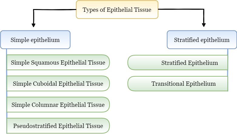
Based on above criteria the epithelial tissue classified into two types:
- Simple epithelium
- Compound epithelium
1. Simple Epithelium
- Simple epithelium consists of a single layer of epithelial cells
- These are direct contact with the basement membrane
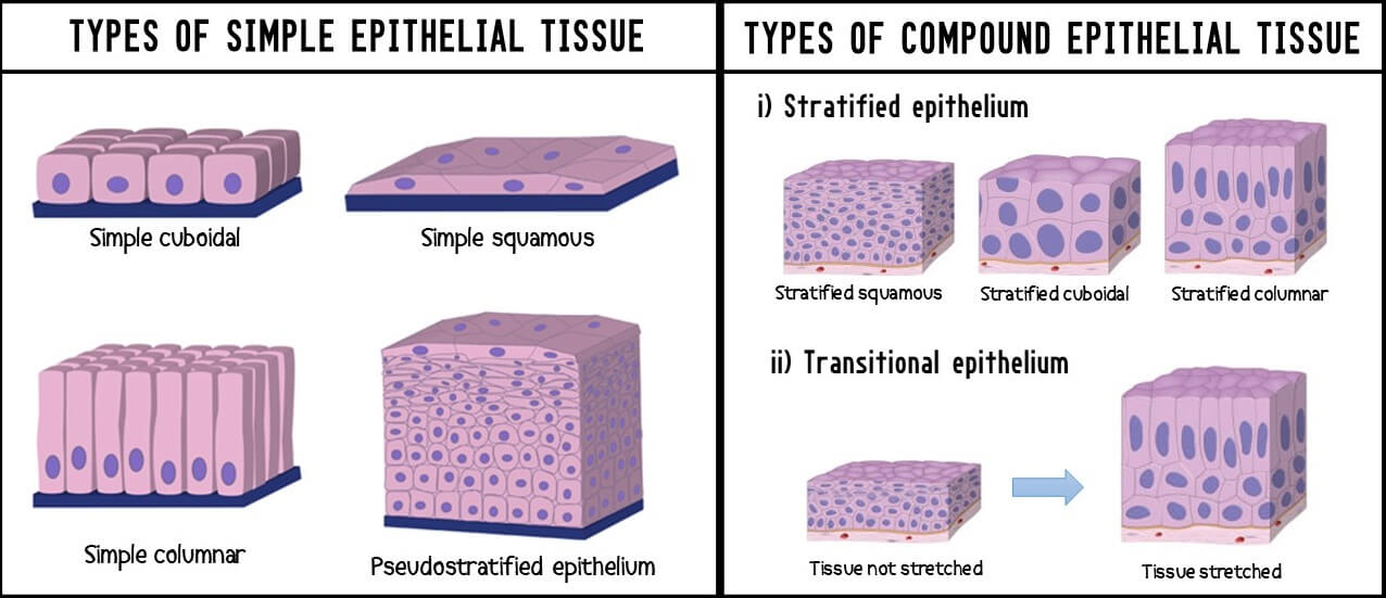
- On the basis of shape and structural modifications of cells the simple epithelium divided into following types:
-
- Simple squamous epithelial tissue
- Simple cuboidal epithelial tissue
- Simple columnar epithelial tissue
- Pseudostratified epithelial tissue
a. Simple squamous epithelial tissue
- Simple = one layer & squamous = flat
- It’s consist of a single layer of thin and flattened cells with nuclei
- On surface view, like floor tiles so also known as pavement epithelium
- Found in lung alveoli, parietal layer of bowman’s capsule of kidney, inner aspect of tympanic membrane, mesothelium and endothelium
- Function is rapid transport of substances, secretion of fluid, diffusion of gases and osmosis
b. Simple cuboidal epithelial tissue
- Simple = one layer & cuboidal = cube
- Made up of single layer of cuboidal shaped cells
- It has round nuclei
- On surface view, cells look like mosaic (hexagonal)
- Found in thyroid follicles, tubules of nephrons, pigmented layer of retina, germinal layer of ovary, inner layer of lens and choroid plexuses of brain
- Function is secretion and absorption
c. Simple columnar epithelial tissue
- Simple = one layer & columnar = rectangular (column)
- These cells having more height than width
- Nuclei are elongated, located in the lower half of cells
- Cells may show some surface modifications
- Common surface modifications of columnar epithelial tissue are microvilli, stereocilia and cilia
- Found in lining of stomach to anus, gall bladder, uterine tube & cavity, central canal of spinal cord
- Function is secretion, absorption and ciliary action
d. Pseudostratified epithelial tissue
- Pseudostratified = false layered (appears to be more than one
- These cells appear layered, but not multi layered
- Nuclei stacked at different levels
- Found in trachea, bronchi, olfactory epithelium, eustachian tube
- Function is secretion and movement of mucus by ciliary action
2. Compound Epithelial Tissue
- It’s consists of many layers, relatively thick
- Cells nearer the surface are flat whereas the deeper are cuboidal and columnar
- On the basis of stretching ability, it is of two types
- Stratified epithelium (non-stretchable)
- Transitional epithelium (stretchable)
a) Stratified epithelium
On the basis of shape of the cells of outermost layer it is of three types
i) Stratified squamous epithelium
- Innermost layer of cells is cuboidal
- Cells of outermost layer are scale like flat cells
- On the basis of presence of keratin protein in the outer most cells this epithelium is of two types
Keratinized stratified squamous epithelium
- Keratin protein is present in scaly cells
- Cells become non nucleated dead cells
- Epidermis of skin, scale, horn, nail, feathers etc
Non keratinized stratified squamous epithelium
- Keratin protein is absent
- Cells are nucleated and living
- Buccal cavity, inner lining of cheeks, lips, hard plate, pharynx, oesophagus & anal canal
ii) Stratified cuboidal epithelium
- Inner most layer is cuboidal
- Outermost layer of cells is cube like, cells are nucleated & living.
- Secretory duct of sweat glands, mammary glands and sebaceous gland, pancreas, salivary glands
iii) Stratified columnar epithelium
- Outermost layer is composed of pillar shaped cells, cells are nucleated. On the basis of presence of cilia this epithelium is of 2 types
(1) Ciliated stratified columnar epithelium. E.g., Buccopharyngeal cavity of frog. Larynx
(2) Non ciliated stratified columnar epithelium. Cilia absent on free end. E.g., Epiglottis
b) Transitional epithelium
- In a relaxed state it’s appears as stratified cuboidal epithelium but tissue is stretched, cells become flattened, giving the appearance of stratified squamous epithelium
- In this epithelium 4-6 layers of cells are present
- Inner most layer of cell is composed of cube like cells
- Middle 2-4 layers are composed of pear shaped
- Outermost 1 or 2 layers are of oval shaped cells
- This different shape of cells appears only in resting stage. When this tissue is stretched, all the cells become flattened
- Found in renal pelvis, ureter, urinary bladder, proximal part of male urethra
- Function the transitional epithelium cells stretch readily in order to accommodate fluctuation of volume of the liquid in an organ
Frequently Asked Questions:-
What are the examples of epithelial tissues?
Epithelial tissue covers external and internal lining of our body. Common example of epithelial tissue is skin. Our skin made up of stratified squamous epithelium. This type of epithelium usually has protective functions
What are the four main functions of epithelial tissues?
Protection, secretion, absorption and filtration is the four main functions of epithelial tissues
Is epithelial tissue avascular?
Yes, that means epithelial tissue lacks blood vessels. They get food and other materials from connective tissue through a basement membrane.
Where is epithelial tissue located?
Simple epithelial tissue found in lung alveoli, parietal layer of bowman’s capsule of kidney, inner aspect of tympanic membrane, mesothelium and endothelium.
You may also like:-
- Epithelial Tissue Structure, Types and Function (With Diagram)
- 3 Types of Simple Permanent Tissue
- Permanent Tissues in Plants & Their Types
- Plant Tissue System Notes
For more detailed information about Structural Organisation in Animals, download now full study material as PDF and if you wana learn more detailed information about Structural Organisation in Animals, visit YouTube Channel.

![[PPT] The living world Class 11 Notes](https://rajusbiology.com/wp-content/uploads/2024/06/PPT-The-living-world-Class-11-Notes-300x169.webp)