Table of Contents
Human Reproduction Notes
In this article we will discuss about Human Reproduction Notes:- Male Reproductive System, Female Reproductive System, Gametogenesis, Menstrual Cycle, Fertilization and Embryonic d Implantation, Pregnancy and Development, Parturition and Lactation
- Reproduction is ability to produce individuals of same species
- Humans are sexually reproducing and viviparous
- Sexual reproduction generally involves 2 parents
- Reproductive events in humans
- Gametogenesis
- Insemination
- Fertilisation
- Implantation
- Gestation
- Parturition
Adolescence
- Time of life when a child reaches reproductive maturity
- Number of changes occurring in the body
- Process of these changes is known as puberty
Reproductive system in human
- Mammalian reproductive system consists of
I) Primary sex organs
- Present at birth
- Also called gonads
- Testes in male and ovaries in female
II) Secondary sex organs
- Develop during puberty
- Prostate, seminal vesicles and penis in male
- Uterus, fallopian tubes and vagina in female
III) External sex characters
- Distinguish two sexes of a species in appearance
- Low pitch voice, beard and narrow hips in male
- High pitch voice, smooth face and broad hips in female
Male reproductive system
- Male reproductive system includes a pair of testes, along with accessory ducts, glands and external genitalia
- Male reproductive system performs two major functions
- Spermatogenesis is formation of sperms
- Transfer of sperm to female reproductive tract
Gonads (testes)
- Located in extra abdominal region
- Testis consists of two flat, oval bodies, one on each side
- Both testes are located in small bag like structure is called scrotum or scrotal sac
- Testis is attached to the scrotum by a band of connective tissue known as gubernaculum
- Temperature of scrotum is 2°C below the body temperature, which is essential for the formation of sperms
- Failure of testis to descend in scrotum from abdominal cavity is called cryptorchidism
Internal structure of testes
- Testis is covered by three layers
- Outer most is tunica vaginalis
- Middle layer is tunica albuginea
- Inner most is tunica vasculosa
- Lobules present in testes are called testicular lobules
- There are about 250 testicular lobules in each testis
- Each lobule filled with 1 to 3 highly coiled seminiferous tubules
- Spermatogenesis occur in seminiferous tubules
- Seminiferous tubules have 2 types of cells
- Male germ cells (Spermatogonia) undergoes spermatogenesis to form sperms
- Sertoli cell (subtentacular cells) provide norishment to germ cell and sperms
- Between the seminiferous tubules, groups of polyhedral cells are found which are called interstitial cells or Leydig cells
- Leydig cells secrete testosterone
Male sex accessory ducts
Rete testes
- Located inside the testis
- Carries sperm from seminiferous tubules
Vasa efferentia
- Connecting link between rete testes and epididymis
Epididymis
- In man it is 6m in length
- Epididymis is divided into three parts
- Upper part: head or caput epididymis (highly coiled)
- Middle part: body or corpus epididymis
- Lower part: tail or cauda epididymis
- Testis and epididymis together are called testicle
- Epididymis function is storage, nutrition and physiological maturation of sperms
- Sperms concentrated and stored in the cauda region until ejaculation
Vas deferens
- Tube like structure emerging from tail part of epididymus
- Opens to ejaculatory ducts
Ejaculatory ducts
- Ejaculatory ducts formed by union of vas deferens and duct of seminal vesicle
- Ejaculatory ducts opens into urethra
Urethra
- Urethra is common passage for urine and sperms
- Originated from urinary bladder
- Open to the exterior through urethral meatus
Male reproductive glands
- Male glands collectively secrete seminal plasma
- Semen = seminal plasma + sperms
Seminal vesicle
- Also called uterus masculina
- It is elongated sac like structure between urinary bladder and rectum
- Secretes seminal fluid
- Seminal fluid is transparent jelly like substance, makes 50-65% of semen.
- Its slightly alkaline (ph 7.3)
- Fructose, ascorbic acid, prostaglandins, bicarbonate
- Fructose is activate sperms
Prostrate gland
- Large gland which lies at the base of the bladder
- It secretes alkaline prostate fluid it is milky, thick and sticky
- It makes about 20-30% part of semen
- Prostate, spermine, citric acid, cholesterol,phospholipids, zinc acid, phosphatease and clotting enzymes
Bulbourethral glands
- Also called cowpers glands
- Pair of glands found in lateral side of urethra
- It secrets transparent slimy, jelly like mucus
- It is slightly alkaline
- Destroys acidity of urethra and cleans it for sperm movement
- It’s also helps in lubrication of penis
External genitalia
Penis
- Penis is male copulatory organ
- Urethra starts at the urinary bladder, pass through the penis and open outside by urethral meatus
- Terminal part of penis is bulging is called glans penis
- Glans penis is covered by movable skin called as prepuce or foreskin
- Inside the prepuce, there are modified oil glands called glands of tyson
- Penis made up of some special type of tissue that helps in erection
- Composed of three longitudinal cylindrical masses of tissues: corpora cavernosa and corpus spongiosum
- External opening of penis is called penile or urethral meatus
FEMALE REPRODUCTIVE SYSTEM
- Female reproductive system consists of pair of ovaries, along with accessory ducts, glands and external genitalia.
- Function of female reproductive system
- Oogenesis is formation of egg
- Allow the entry of sperms
- Site for fertilisation
| FEMALE REPRODUCTIVE SYSTEM | |
| Gonads | Pair of ovaries |
| Accessory Ducts | Pair of oviduct, Uterus and vagina |
| External genitalia | Mons pubis, Labia majora, Labia minora, Hymen and Clitoris |
| Glands | Bartholin gland, Skene gland and mammary glands |
OVARIES
- Ovaries are primary sex organ in female
- Located one on each side of lower abdomen
- Ovary connected to pelvic wall and uterus by ligaments
- Ovary length is 2 to 4 cm
- Function of ovary
- Oogenesis
- Secretion of hormones (Estrogen, Androgen, Progesteron and inhibin)
Ligament of ovary
- Ovarian ligament is fibrous ligament that connects ovary to side of the uterus
- Broad ligament helps to support the uterus and maintain its position in the pelvic cavity
- Mesovarium is the portion of the broad ligament. it suspends the ovaries
Internal structure of ovary
- Ovary is covered by layer of cubical epithelium called germinal epithelium
- Beneath this epithelium, tunica albuginea is present
- Inner region of ovary is called stroma, its divided into 2 zones
- Cortex: Peripheral, dense and contain ovarian follicles
- Medulla: Inner, abundant blood vessels and nerve fibres
- Follicular cells
- They are supporting cells
- Surrounded the oocyte within the follicle
- Follicular cells produces hormones
- Maturation of follicle is called folliculogenesis
Folliculogenesis
Primodial follicles
- Developed in fetal stage
- Composed of primary oocyte surrounded by squamous epithelium
Primary follicles
- Developed after puberty
- Composed of primary oocyte surrounded by cuboidal epithelium
Secondary follicles
- Composed of primary oocyte surrounded by many layer of follicular cells known as granulosa cells
Graafian follicles
- Composed of secondary oocyte surrounded by many layer of follicular cells
- Following layers of cells are observed from the oocyte
- Corona radiata: Immediately around the zona pellucida
- Corona radiata: Cluster of cell surround the oocyte
- Granulosum: present just after the cumulus cells. its secrete liquor fluid of the antrum
- Teca interna: Spindle shaped cells around the granulosa cells
- Teca externa: Outer most layer of loose connective tissue
Ovalation
- Release of mature oocyte from the ovary to the fallopian tube is called ovulation
Process of ovulation
- Mature follicle protrudes from the ovary. this condition is called stigma
- Increase secretion of liquor folliculi
- Folicular wall become thin and rupture
- Relase of oocytes
Empty ovarian follicles
Corpus hemorrhagicum
- During ovulation blood vessels ruptured which cause bleeding inside the follicle
- The follicle looks like red mass, hence known as corpus hemorrhagicum
Corpus luteum
- The granulosa cell and theca interna cell enlarge and hypertrophied
- Accumulate yellow pigment (lutein)
- The mass now become yellow. This mass is called corpus luteum
Corpus albicans
- After the regression of corpus luteum the mass is replaced by white tissue
ACCESSORY DUCTS AND UTERUS
FALLOPIAN TUBES
- Also known as Oviducts or uterine tube
- Fallopian Tube carry eggs from the ovaries to uterus
- Oviduct is differentiated into three parts
Infundibulum
- Funnel shaped end of fallopian tube is called infundibulum
- Fimbriae, found on the lateral end of each tube, are fringe like protrusions
- It helps in collection of ovum after ovulation
Ampulla
- Infundibulum leads to ampulla
- Ampulla is the wider and longest part of oviduct
- Fertilization of ovum takes place
Isthmus
- It is last part of the oviduct
- Isthmus opens to uterus
UTERUS OR WOMB
- Uterus look like unverted pear shaped
- Located in the pelvic region between urinary bladder and rectum
- It is attached to pelvic wall by ligaments
- Major portion of uterus is the body
- Rounded region above to body is fundus
- Uterus opens into vagina through a narrow cervix. Its cavity is termed as cervical canal
- Birth canal = Cervical canal + Vagina
- Uterus is the site for implantation
Uterus wall layers
- Uterus walls is composed of three layers
Perimetrium
- External thin layer
- Made up of loose connective tissue
Myometrium
- Midlle thick layer of smooth muscles
- Myometrium exhibits strong contraction during delivery
Endometrium
- Inner glandular layer
- Endometrium is composed of two layers: basilar and functional
- Basilar layer is permanent, vascular and very thin
- Functional layer of endometrium is regenerated and lost during each menstrual cycle
VAGINA
- Vagina is muscular tube about 810cm long
- Vagina is female copulatory organ
- The vagina receives sperm from the male penis during sexual intercourse
- Opening of the vagina in young females is partially closed by thin membrane called hymen
- During reproductive life the vagina contains Lactobacillus acidophilus
- These bacteria produce lactic acid from glycogen. it keeps vaginal pH between 4.9 to 3.5
- This acidity helps to prevent vaginal infections
EXTERNAL GENITALIA
- Accessory structure of female reproductive system that are external to the vagina
- Vulva includes the mons pubis, clitoris, labia majora, labia minora and Bartholin’s gland
- Mons pubis: Its fatty tissue cushion which is covered by skin and public hair
- There are 2 pairs of lips like tissue around vagina
- Labia majora: Outer fleshy folds surrounding the vagina
- Labia minora: Inner fleshy folds located under labia majora
- Clitoris: Small finger-like structure located at the upper meeting point of two labia minora
FEMALE ACCESSORY GLANDS
Bartholins gland
- Pair of glands situated on each side of vaginal opening
- Secretion of Bartholin’s gland help in lubrication during copulation
Skene’s glands
- Pair of glands situated around the lower end of the urethra
- They secrete a lubricating fluid
Mammary gland
- Mammary Gland are found in all female mammals
- Mammary Gland functional only in females
- Overgrowth of breast in male is called Gynaecomastia
- Externally, each breast has raised nipple, which is surrounded by a circular pigmented area called areola
- Mammary glands composed of glandular tissue and fats
- Mammary gland function is regulated by hormones
- Estrogen – Stimulate the development of glandular tissue
- Progesterone – Stimulate the development of the duct system
- Prolactin – Stimulate the production of milk
Internal structure of mammary gland
- Adult female breast contains 15 to 20 lobules of glandular tissue
- Each lobule divided into numerous alveoli
- Alveoli is group of cells having lumen within
- Alveoli secrete milk, its stored in the cavities of alveoli
- Alveoli open in mammary tubules which join to form mammary duct
- Several mammary ducts join to form ampulla
- Mammary ampulla continues as lactiferous duct and open at surface of nipple
GAMETOGENESIS
- Gametogenesis is the process of gamete formation
- Gametes are formed from germ cells of gonads
- There are two types of gametogenesis
- Spermatogenesis
- Oogenesis
SPERMATOGENESIS
- Spermatogenesis is the process of producing sperms
- Occurs in seminiferous tubules at the time of puberty and continues throughout life
- Hormones regulates spermatogenesis
- Gonadotropin releasing hormone (gnRH): secrete from hypothalamus at puberty
- Luteining hormone (LH): stimulate leydig cells and synthesis testosteron
- Follicle stimulating hormone (FSH): stimulate sertoli cells
- Stages of spermatogenesis
- Formation of spermatids: germ cell produce spermatids
- Spermiogenesis: spermatids transform into sperm
Stages of spermatogenesis
I) Formation of spermatids
- Spermatid formation occurs by 3 phases
Multiplication
- Spermatogonia present inside the wall of seminiferous tubules
- Spermatogonium divide mitotically to increase in number
Growth
- Some spermatogonia stop dividing
- Some spermatogonia actively grows into primary spermatocyte
- That growing cells obtain nourishment from sertoli cells
Maturation
- Primary spermatocyte complete meiosis i to produce 2 cells called secondary spermatocytes
- Both secondary spermatocytes undergo meiosis ii to produce 2 spermatids each
II) Spermiogenesis
- Spermatids are non motile
- Each spermatid is transformed into sperms
- Spermatids head remain attach to sertoli cells, tail hanging in lumen of seminiferous tubules
- Sperms are finally released from seminiferous tubules by the process called spermiation
Changes during spermiogenesis
- Condensation of nucleus
- Spermatid length increases
- Centrioles rearranged
- Golgi body breakdown to form acrosome
- Mitochondria coiled
Structure of sperm
- Human sperm was first seen by hamm and leeuwenhoek
- Human sperms move at speed of 3mm/min through female genital tract
- The spermatozoa has four parts: head, neck, middle piece and tail
Head
- The head contains two main parts
- Nucleus: elongated
- Acrosome: bag like structure filled with lytic enzymes called sperm lysin
Neck
- Neck is very short containing proximal and distal centriole
- Two centrioles arranged one after the other behind the nucleus
Middle piece
- Middle piece contains spiral rows of mitochondria
- Provide energy and strength to the sperm for locomotion
Tail
- Vibrating posterior portion of sperm which helps in swimming
- Tail is made up of axial filament and small amount of cytoplasm
OOGENESIS
- It is type of gametogenesis in females
- Oogenesis is the discontinuous process
- This process begins before birth, stops in mid and resumes after puberty
- Hormones regulates oogenesis
- Luteinzing hormone (LH): stimulate the ovulation and maintaining corpus luteum
- Follicle stimulating hormone (FSH): stimulate the follicular cells for maturation
- Oogenesis occurs through the following phases
- Multiplication phase
- Growth phase
- Maturation phase
Phases of oogenesis
I) Multiplication phase
- In this stage germs cells repeatedly divide by mitosis
- Result of mitosis produce large number of egg mother cells or oogonia
- This happens 3 to 8 months of gestation in embryo
- Millions of oogonia formed within ovaries
- This process completes in embryo stage
II) Growth phase
- Growth phase is longest phase in oogenesis
- In this process oogonia grow in size and form primary oocytes
- During growth phase size of cells increases many times
- Primary oocytes enter into meiosis i and get temporarily arrested at prophase i diplotene stage
- Primary oocytes then get surrounded by layer of granulosa cells to form primodial follicles
III) Maturation phase
- In this stage primary oocytes under go meiosis i to produce two haploid cells
- Larger one called secondary oocyte and smaller one called polar bodies
- Secondary oocyte proceeds meiosis ii, only up to metaphase ii and division is arrested at this stage
- Then secondary oocyte is shed from the graffian follicle and ovary
- Secondary oocyte goes inside the fallopian tube and wait for sperm
- If the secondary oocytes doesn’t receive sperm, it disintegrates and shed off in menstrum
- If sperm meet the secondary oocytes fertilisation gets started
- Meiosis ii restart and completion with help of spermatozoa
- End of meiosis ii two unequal cells formed, large cell is ovum and small cell is polar body
Structure of human egg
- Human egg is largest cell in the body
- Human egg also known as mature ovum or secondary oocyte
- Mature ovum covered with 3 distinct membranes in human
- Corona radiata: made up of follicular cell
- Zona pellucida: thick non cellular membrane and developed from follicular cells
- Vitelline membrane: forming surface of cytoplasm
- Cytoplasm have 2 zones
- Exoplasm: peripheral and less dense
- Ooplasm: inner and contain nucleus, organelles, yolk
CLASSIFICATION OF EGGS
I) On the basis of amount of yolk
Microlecithal egg
- The amount of yolk is much less than the amount of cytoplasm
- Examples: mammalian eggs
Mesolecithal eggs
- The amount of yolk is moderate
- Examples: eggs of amphibia
Macrolecithal eggs
- Eggs are with large amount of yolk
- Examples: insects egg and birds
II) On the basis of distribution of yolk
Isolecithal eggs
- Yolk is evenly distributed in these eggs
- Example: mammalian eggs
Telolecithal eggs
- Yolk is concentrated in one part of the egg
- Example: eggs of amphibia
Centrolecithal eggs
- Yolk is located in the centre and surrounded by cytoplasm
- Example: insects egg
III) On the basis of shell
Cleidoic eggs
- Eggs surrounded by hard shell
- These eggs have more amount of yolk
- Example: birds and reptiles eggs
Non cleidoic eggs
- Eggs are not surrounded by hard shell
- Examples: mammals and amphibians
MENSTRUAL CYCLE
- Process of bleeding through vagina due to breakage of endometrium is called menstruation
- In this cycle female body prepares itself for pregnancy
- If pregnancy does not occur then the body aborts all preparation
- In healthy female menstruation occurs at average interval of about 28 days
- Menarche: first menstruation that begins at puberty
- Menopause: menstrual cycle stop at about 50 years of age
- Menstrual cycle has four main phases
- Menstruation phase
- Follicular phase
- Ovulatory phase
- Luteal phase
Phases of menstrual cycle
I) Menstruation phase
- Days 15 of the cycle are known as menstrual phase
- During menstrual phase, the layer of endometrium gets shed off
- Due to breakdown of endometrial lining of uterus, bleeding continues upto 4-5 days
- Blood clotting doesn’t occur due to presence of fibrinolytic enzymes
- In every cycle 40-80 ml blood loss
Hormonal change
- Progesterone are low
- FSH increase due to negative feedback of low level of progesterone
II) Follicular phase
- Uterine bleeding is stopped
- Follicle grows, matures and secretes estrogen during this phase
- Endometrium line start to repair
Hormonal change
- High estrogen is stimulate pituitary gland for decrease FSH and increase LH secretion
- This abrupt rise in LH concentration in blood is called LH surge
III) Ovulatory phase
- Occurs around 14th day
- Rupture of graafian follicle and release of an ovum
Hormonal change
- Fall in FSH level and arise in LH
- Increased concentration of LH causes the graafian follicle to rupture
IV) Luteal phase
- The ruptured graafian follicle transforms into corpus luteum
- Corpus luteum secretes progesterone and some estrogens
- It fertilization occurs
- Corpus luteum persists
- If fertilization does not take place
- Corpus luteum remains functional for 10 days
- After 10 days, begins to degenerate into a corpus albicans
- This causes disintegration of the endometrium leading to menstruation
Hormonal change
- If ovum is not fertilized, the higher level of progesterone inhibits the production of LH
- Withdrawal of LH causes regression of corpus luteum and fall in progesterone level
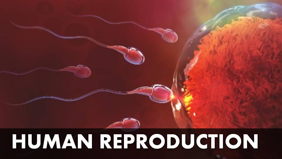
FERTILIZATION
- Process of fusion of male and female gamtes to form zygote is called fertilization
- Fertilization normally occurs in ampulla region
- Fertilization only occur if ovum and sperm are transported at the same time in ampulla
- Fertilization involves the following steps
- Entry of sperm
- Fertilizin and antifertilizin reaction
- Release of sperm lysin
- Fertilization cone
- Completion of meiosis ii
- Cortical reaction
- Fusion of gametes
Entry of sperm
- In male single ejaculation contain 3ml of semen, its deposited in vagina
- Nearly 100+ sperms reach fallopian tube
- Capacitation is the preparation of sperm to fertilize the ovum
- Removal of sterol and glycoprotein and ca2+ ions taken
Fertilizin and antifertilizin reaction
- Fertilizin is a protein secreted from egg cytoplasm
- Antifertilizin is a protein present in plasma membrane of the sperm
- Fertilizin acts as receptor for antifertilizin
- Reaction between fertilizin and antifertilizin makes the sperm more capable of fertilizing the egg of same species
Release of sperm lysin
- Corona digestive enzyme: digest the cells of corona radiata
- Acrosin: digest zona pellucida
Fertilization cone
- Acrosome of sperm touches surface of egg, the cytoplasm of egg bulges forward forming receptive cone or fertilization cone
- Receptive cone is the region where sperms enters the egg
Completion of meiosis ii
- Metaphase factors are deactivated
- Anaphase promoting factors are activated
- Secondary oocyte undergoes meiosis ii, resulting in formation of ovum and second polar body
Cortical reaction
- Entry of other sperm is prevented by development of fertilization membrane
- Massive exocytosis of cortical granules to diffuse and alter the structure of zona pellucida.
- In human both head and tail of sperm enter the cytoplasm of oocyte
Fusion of gametes
- Nucleus of sperm absorb water from egg cytoplasm and becomes enlarged
- Enlarged male nucleus is called male pronucleus
- Nuclei of both gametes fuse to form zygote
- Fusion of gametic pronuclei is called karyogamy
- Mixing of two sets of chromosomes of two gametes is called amphimixis
Significance of fertilization
- Help to restore the total number of chromosomes in the organism
- Fertilization combines the characters of two parents. This brings genetic variations
- It determines the sex of the embryo in human
MORULA FORMATION
- After fertilization, the zygote continue to divide by rapid mitotic cell division called cleavage
- Cleavage in fertilized egg starts in fallopian tube after fertilization
- The large zygote divide into many smaller daughter cells called blastomeres
- After 4-5 cleavages approximately 16-32 cells formed
- There is no growth phase between cleavages
- This solid ball of cells called morula
Cleavage
- Cleavage also known as cellulation and segmentation
- The term was given by von baer
- After fertilization, zygote continue to divide by rapid mitotic cell division called cleavage
Types of cleavage
- Determinate cleavage: fate of the cells being set early in the embryo development
- Indeterminate: each cell has the potential of developing into a complete organism
Stages of morula formation
Zygote to 2 cell stage
- Occurs 24-30 hours after fertilization
- Unicellular zygote changes into two blastomeres
- Occurs in the upper portion of fallopian tube
2 cell stage to 4 cell stage
- Occurs after 1st cleavage
- Larger cell divide and form 3 cell stage
- Then later smaller cell also divide and form 4 cell stage
4 cell stage to 8 cell stage
- 3rd cleavage occurs
- Cleaved cells pushed from fallopian tube towards uterus
8 cell stage to morula
- At the end of 4th cleavage of fertilization
- The solid ball of 16-32 cells formed
- This solid ball like structure is called morula
- No change in overall size from zygote to morula
BLASTULATION
- Blastula formation is called blastulation
- Morula changes to blastula due to rearrangement of blastomeres
- Blastula is typically a hollow, multicellular ball like cells produced at the end of cleavage
- Fluid filled cavity of blastula is called blastocoel
- Blastocyst is formed, zona pellucida becomes thinner and finally disappears
- By this time the developing embryo reaches the uterus and implants into the uterine wall
Structure of blastocyst
- Blastocyst has 3 parts: trophoblast, inner cell mass and blastocoel
Trophoblast
- Single outer layer of large flat cells
- Helps in absorbing nutrition fro the embryo
- Later form into chorion, amnion and part of placenta
Embryoblast
- Inner cells mass forms the body of embryo
- Embryo develops from embryoblast
- Thin layer of trophoblast that overlies the embryoblast
Blastocoel
- Fluid filled cavity which helps in rapid expansion of blastocyst
IMPLANTATION
- Attachment of the blastocyst to the uterine wall
- Implantation takes place about seven days after fertilisation
- Zona pellucida help to prevent the implantation of blastocyst at an abnormal site
- Upon implantation, the endometrium undergoes changes referred to as the decidual cell reaction
- Blastocyst attaches itself by embryonic pole, close to the endometrium
- Increases the permeability of blood vessels and wall of endometrium
- Trophoblast cells of blastocyst have power to stick to the uterine wall
- Endometrium get disturbed by finger like structure is called chorionic villi
- Chorionic villi penetrate by lytic or mechanical way
- By the end of 10th day, whole embryo is deeply embedded into the endometrium, completing implantation process
GASTRULATION
- Process of single layered blastula is transformed into gastrula
- Gastrula covered with three layers called germ layers
- Germ layers are give rise to specific parts of the organism
Ectoderm
- Outermost layer of developing embryo
- Give rise to integumentary system (the outer skin, nails and hair) as well as the nervous system (central and peripheral system)
Endoderm
- Innermost layer of developing embryo
- Gives rise to epithelial layer of the digestive tract, lungs, pancreas, bladder, liver as well as the thyroid gland, parathyroid gland and thymus
Mesoderm
- Middle layer of developing embryo
- Give rise to musculoskeletal system (bone, cartilage, skeletal muscle, cardiac muscle, smooth muscle), cardiovascular system (the heart and blood vessels), excretory system (kidneys) and reproductive system (gonads)
Formation of layers by gastrulation
Formation of endoderm
- Blastocyst grows in size by obtaining nutrition from the uterus
- Some cells separate from embryonic knob to form endoderm in blastocoel
- Remaining inner cell mass forms embryonic disc
Formation of mesoderm
- Mesoderm is formed from the caudal margin of the embryonic disc.
- Prior to this the existing cells undergo rapid division and a mass of cells detach from the embryonic disc to form mesoderm.
Formation of ectoderm
- After the separation of mesoderm, the remaining cells of the embryonic disc form the ectoderm layer
- In this manner the three germ layers such as ectoderm, mesoderm and endoderm are formed.
PLACENTA
- Placenta is structural and functional connection between foetus and maternal tissue
- Placenta is found in all viviparous animals
- Placenta is formed of tissues from mother and foetus. So called as fetomaternal organ
- Chorionic villi and uterine tissue become interdigitated with each other
Placenta has two components
- Fetal part – develops from chorion
- Maternal part – develops from functional layer of decidua basalis
Umbilical cord
- Umbilical cord connecting the foetus with the placenta
- Umbilical cord contain two umbilical arteries and one vein
- Blood vessels in umbilical cord are surrounded by wharton’s jelly
- Placenta and umbilical cord are transport system for substances between mother and fetus
Functions of placenta
- Exchange of materials between foetal and maternal blood
- Placenta also serves as a repiratory medium for exchange of o2 and co2
- The nitrogenous and metabolic waste from foetus are released into blood of mather
- It also produces hormones
- Human chorionic gonadotropin (HCG)
- Human placental lactogen (HPL)
- Progestogens and estrogens
- Provides passive immunity antibodies
- Harmful drugs and chemicals cross placenta and cause foetal deformity
- Some viruses may cross the placenta and cause infection to foetus
EXTRA EMBRYONIC MEMBRANES
- Central part of blastula gives rise to embryo
- Peripheral portion of blastula does not take part in embryo formation
- The peripheral part is known as extra embryonic region or trophoblasts
- This extra embryonic region takes part in formation of certain membranes called extra embryonic membranes
- Extra embryonic membranes are four types: chorion, amnion, yolk sac and allantois
- On the basis of amnion two groups of vertebrates are categorised
- Amniota – this group of animals have amnion in the embryos. E.g. reptilia, aves and mammalia
- Anamniota – this group of animals devoid of amnions in their embryos. E.g. cyclostomata, pisces and amphibia
Four types of extra embryonic membranes
Chorion
- Chorion is the outer foetal membrane
- Chorion is the name given to trophoblast after formation of embryo
- Chorion helps in gaseous exchange
- Acts as extra embryonic lungs in reptiles, birds and prototherians
Amnion
- A thin protective membrane that surrounds the embryo
- Amniotic cavity filled with fluid called amniotic fluid
- Fetal cells are present in amniotic fluid
- Embryo in this stage is called as foetus remains hanging in amniotic fluid
- Amnion well developed in amniote (mammals, birds, reptiles)
Yolk sac
- Initially the size of yolk sac is larger as compared to that of embryo
- Behaves like extraembryonic gut
- In human it is vestigial and after 8 weeks size reduced
- Well developed in bird and reptiles
Allantois
- Allantois is a hollow sac like structure
- In human allantois does not function
- Allantois works well in reptiles, birds and prototherians
- Acts as extra embryonic kidney
- Allanotis stores nitrogenous wastes
PARTURITION
- Parturition means birth of the baby
- The average duration of human pregnancy is about 9 months called as the gestation period
- During parturition, uterus contracts to push the fetus towards cervix
- This contraction continues until the fetus comes down the birth canal
- After parturition, uterus releases the placenta and it passes out immediately after the fetus is born
Mechanism of parturition
- Fully developed foetus and the placenta gives signals for parturition
- Initiation of mild uterine contractions called foetal ejection reflex
- Due to mild uterine contractions, the oxytocin release from pituitary
- Oxytocin causes stronger uterine contractions
- Due to these stronger contractions, the foetus starts moving towards the vagina
- The labour pain during child birth is due to this hormone.
- After parturition oxytocin stimulates milk ejection
- Relaxin hormone is secreted by placenta and ovary.
- This hormone relaxes the pubic symphysis
Three stages of parturition
Dilation stage
- Cervix and vagina gets dilated
- Uterine contractions begin from top, forcing the baby towards the cervix
- Oxytocin induced uterine contractions
- As the baby is pushed down in the uterus, its head comes to lie against cervix
- It ends in rupturing of amniotic membrane of foetus
Expulsion stage
- This stage starts at full dilation and continues until birth
- The foetus passes out through cervix and vagina with head in forward direction
- This stage ends with the childbirth and the umbilical cord is clamped
Placental stage
- After the delivery of the baby the placenta separates from the uterus and is expelled out
LACTATION
- Lactation is process of milk secretion from the mammary glands after childbirth
- Mother milk provides nutrition and immunity to the young one
- The secretion of milk from alveoli of mammary glands starts from end of pregnancy period
- The ejection of milk starts after child birth
- Lactation is complex neuro endocrine process
- Lactation is divided into four phases
- Preparation of breasts
- Secretion of milk
- Ejection of milk
- Maintenance of lactation
Stages of lactation
Mammogenesis
- Mammary gland growth occurs
- The size and weight of breast increases
- Generally, a female is ready to produce milk during the fifth or sixth month of her pregnancy
- Progesterone influences growth in size of alveoli and lobes
- Estrogen stimulates the milk duct system to grow and differentiate
Lactogenesis
- Alveoli of mammary glands secrete milk
- Prolactin secretion is stimulated when estrogen and progesterone levels are low
- Placenta is responsible for progesterone production. After parturition, placenta loss so progesterone decreases suddenly
- Low level progesterone stimulates anterior lobe of pituitary to secrete prolactin
- Now, prolactin stimulates milk secretion
Galactokinesis
- Ejection of milk from the alveoli of the mammary glands
- Milk ejection is under the control of oxytocin
- Oxytocin is released from posterior pituitary gland during nipple stimulation or sensory stimulation
- Oxytocin causes ejection of milk from alveoli by contraction of muscles around mammary gland
Galactopoiesis
- Galactopoiesis is the maintenance of lactation once lactation has been established
DISORDERS OF REPRODUCTIVE SYSTEM
Oligospermia
- Oligospermia is a male fertility issue characterized by a low sperm count
- World health organization (who) classifies sperm counts
- Mild oligospermia is 10 to 15 million sperm/ml
- Moderate oligospermia is considered 5 to 10 million sperm/ml
- Severe oligospermia is diagnosed when sperm counts fall between 0 and 5 million sperm/ml
Azoospermia
- Azoospermia means there’s no sperm in a man’s ejaculate
- Its causes include a blockage along the reproductive tract, hormonal problems, ejaculation problems or issues with testicular structure or function
- Many causes are treatable and fertility can be restored
Cryptorchidism
- Cryptorchidism is the absence of one or both testes from the scrotum
- It is the most common birth defect of the male genital tract
Gynecomastia
- Gynecomastia is enlargement of breast size in males
- Its happen due to reduced male hormones (testosterone) or increased female hormones (oestrogen)
Hysterectomy
- Hysterectomy is an operation to remove the uterus
- This surgery may be done for different reasons
- Once you’ve had a hysterectomy, you’ll stop having menstrual periods
Oophorectomy
- Oophorectomy is surgical procedure to remove one or both of ovaries
- When you have one ovary removed, it’s called unilateral oophorectomy. Removal of both ovaries is called bilateral
Ectopic pregnancy
- Ectopic pregnancy occurs when a fertilized egg implants and grows outside the uterus
- Ectopic pregnancy most often occurs in a fallopian tube
Teratogens
- Some substance that can disturb the development of an embryo or fetus
- Teratogens may cause a birth defect in the child

![[PPT] The living world Class 11 Notes](https://rajusbiology.com/wp-content/uploads/2024/06/PPT-The-living-world-Class-11-Notes-300x169.webp)